
Musculoskeletal Ultrasound for Achilles Tendonitis in Towcester
Book a musculoskeletal ultrasound for Achilles tendonitis in Towcester. Get clear answers, prompt diagnosis, and a practical plan to get back to normal.
A sore Achilles can make everyday plans feel uncertain, especially when you’re unsure whether to keep walking or running. Musculoskeletal ultrasound for Achilles tendonitis in Towcester provides moving images of the tendon and nearby soft tissue, helping clinicians investigate what may be contributing to your symptoms.
The scan is only one part of the picture. When the discomfort began, which activities bring it on and how it affects your day all help put the images in context. A scan alone cannot show how your heel responds on stairs, during a walk or after sitting at your desk.
This article explains how ultrasound assessment works, what changes in the tendon and surrounding tissue may look like, and how anatomy helps make sense of the findings. You’ll also learn what to discuss during an assessment and how the results can help shape practical next steps towards your usual activities.
Key Takeaways
- Notice where your pain sits and when it flares, these details help make scan findings more meaningful.
- Learn what the scan can reveal about the tendon itself and the tissue around it.
- Understand why a suspected tear needs careful assessment rather than guesswork based on symptoms alone.
- Use the assessment findings to shape a practical plan for returning to the activities that matter to you.
Musculoskeletal ultrasound assessment of Achilles tendinopathy
Ultrasound provides moving images of the Achilles tendon and nearby soft tissue. Clinicians interpret these alongside your symptoms and physical examination.
Tendon thickening and vascularity
Tendinopathy can make the tendon thicker and darker on imaging. Doppler ultrasound may show increased blood flow. A normal tendon is commonly described as 4 to 6 mm thick, while symptomatic tendons frequently measure more than 7 mm. These features add context, but thickness alone cannot confirm the cause of pain.
Insertional symptoms and assessment
Insertional tendinopathy affects the lowest 2 cm of the tendon, where it joins the heel bone. Pain at the back of the heel, particularly with shoe pressure or ankle movement, can prompt closer assessment of this area. Identifying the pain location helps the clinician interpret the scan alongside your symptoms.
Tendon movement and continuity
During a dynamic assessment, the clinician watches the tendon as your ankle moves. This can show how the tendon glides and whether its fibres remain continuous. A visible gap or restricted movement may help explain your symptoms, but scan findings must be considered alongside your examination.
Scan procedure, preparation and results
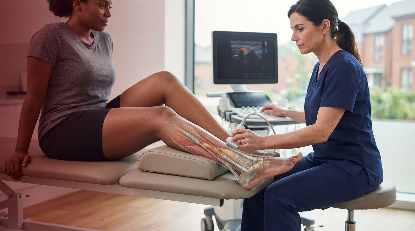
You usually lie face down, with your foot supported or hanging freely, while gel and a probe are used to examine the tendon. Special preparation is generally unnecessary. Ultrasound is non-invasive, but image quality and interpretation can limit what it reveals.
A GP referral is not universally required for private musculoskeletal ultrasound in the UK. NHS scans are usually arranged through a GP or another clinician, when examination suggests imaging may help guide care; local referral pathways determine access. There is no single results timeframe: it depends on the service’s reporting process. Findings may be explained as part of your assessment or sent to the referring clinician. A plan may also include preventative care.
Ultrasound differentiation of tendinopathy, paratendinitis and tendon tears
Ultrasound can help identify which part of the Achilles region appears affected. The key distinction is whether changes are within the tendon, around it or interrupting its fibres.
Insertional changes and retrocalcaneal bursitis
At the heel attachment, a scan can assess the tendon and retrocalcaneal bursa as separate structures. The bursa is a fluid-filled space between the Achilles tendon and heel bone. Identifying whether a change lies within the tendon or in this neighbouring space helps clarify which tissue may be involved. This matters because similar discomfort can come from different structures.
Symptoms and tendon locations
Achilles tendinopathy commonly affects the middle portion of the tendon or its attachment to the heel bone. Tears typically occur 2 to 6 cm above that attachment, so the scan examines this region carefully when injury is suspected. For example, a sudden loss of push off strength after an abrupt injury may lead the clinician to assess fibre continuity closely, rather than focus only on the painful spot.
Ultrasound findings in tendinopathy
Tendinopathy affects the tendon itself, while paratendinitis involves the tissue around it. A tear may appear as disrupted fibres; a complete acute rupture may show retracted ends and a blood-filled gap. These patterns can help distinguish conditions, but images must be interpreted alongside how the problem began and what you can do during examination. A scan supports clinical reasoning rather than establishing a diagnosis on its own.
Ultrasound can distinguish tendinopathy from paratendinitis and partial or complete tears. Retrocalcaneal bursitis is inflammation of the bursa between the Achilles tendon and heel bone. In insertional Achilles tendinopathy, lowering the heel over a step during tendon loading may aggravate symptoms as the calcaneus presses against an inflamed bursa or tendon. Approximately 24% of people suffering from heel pain were diagnosed with insertional Achilles tendinopathy; roughly one-third of people with Achilles tendon pain have insertional Achilles tendinopathy. In the study, longitudinal-plane Achilles tendon echogenicity averaged 69.6/255 in symptomatic tendons and 77.0/255 in asymptomatic tendons.

Achilles tendon anatomy and ultrasound appearance
The Achilles tendon connects the calf muscles to the heel bone, transferring force when you walk, climb or run. Understanding its normal structure helps make ultrasound findings more meaningful, especially when symptoms are persistent or affect sport.
The tendon spans approximately 15 cm, running from the calf muscle towards its insertion on the heel bone. On ultrasound, an intact tendon has an organised fibre pattern, visible in long axis and cross section. One set of stated normal measurements is 4.3 mm thick by 14.3 mm wide. Measurements vary with the scan plane and the person, so a single figure cannot explain pain by itself.
In a long-axis image, the fibres appear as parallel lines along the tendon. In a transverse image, they form a compact pattern across it. The clinician compares both views and may assess the symptomatic side alongside the other tendon, helping distinguish a local change from normal variation.
Imaging also shows where a finding is located. A change near the heel insertion differs from one higher up the tendon, even if both appear thickened. For example, findings for a runner with pain at the heel attachment should be considered alongside footwear pressure and loading history, not judged by tendon size alone.
Ultrasound findings can guide the next clinical conversation, but a scan does not measure strength or explain every cause of pain. Your symptoms, movement and response to loading complete the picture. If Achilles pain is affecting your activity, arrange a musculoskeletal assessment to discuss suitable imaging and next steps, while podiatry practices such as Active Foot Care can assist with broader foot pain and mobility management.
RED Physiotherapy: Physiotherapy
RED Physiotherapy can help you make sense of Achilles symptoms and turn assessment findings into a practical physiotherapy plan. If heel pain is limiting walking, running or work, an assessment can explore how your symptoms affect movement and daily activities, then identify suitable next steps.
Care is tailored to your goals, whether you hope to return to sport or feel more comfortable day to day. Physiotherapy may include hands-on treatment and a clear plan for progressing activity, rather than exercises without context. Contact RED Physiotherapy to discuss an assessment and your next steps.
Take your next step towards confident movement
A scan can clarify what may be happening, but your next step should also suit your symptoms, routine and goals. If you’re considering musculoskeletal ultrasound for Achilles tendonitis in Towcester, think about what you want to return to, whether that’s comfortable daily walking or sport. This gives your assessment a practical focus.
RED Physiotherapy provides physiotherapy to help you understand your options and plan appropriate progress. Sports massage may also support your wider care, while women’s health physiotherapy addresses needs specific to pelvic health and movement. The right approach depends on the concern you want help with.
Ready to talk through your Achilles symptoms and possible next steps? Contact RED Physiotherapy about your Achilles, and take a considered step towards moving with greater confidence.
Frequently Asked Questions
How accurate is ultrasound for diagnosing Achilles tendonitis compared with MRI?
There isn’t one accuracy figure that applies to every Achilles problem. Ultrasound provides a real-time view of the tendon and can assess movement, while MRI gives a different view of the area. Which is more useful depends on the clinical question, such as suspected tendon changes or a tear. Your symptoms and examination help determine how imaging findings should be interpreted.
Can ultrasound distinguish Achilles tendinopathy from a tendon tear or bursitis?
Yes, ultrasound can help distinguish these conditions by showing which tissue appears affected, though findings need clinical interpretation. For example, pain after a sudden injury accompanied by difficulty pushing off needs prompt assessment for a tear. Pain around the heel may instead involve the bursa or tendon attachment. Tell the clinician how your symptoms began and which movements make them worse, as this context helps interpret the scan.
Tags:
About the Author
Red Physiotherapy Team
Content Team
Health and wellness content from the RED Physiotherapy team.